
Treatment
Evaluation & Diagnosis
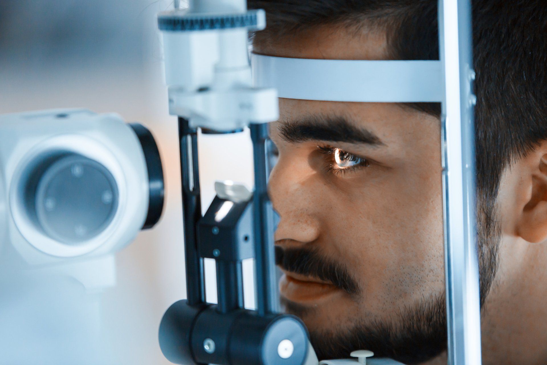
Our medical staff can often evaluate, test, diagnose and treat you. As a result, a comprehensive evaluation, with or without treatment, could last anywhere from two to four hours.
An ophthalmic technician will ask all first-time patients to provide a thorough medical history, including all medications. On each visit, we will perform a vision test and a measurement of the intraocular pressure, followed by dilation of the eyes. After a wait to allow your pupils to dilate adequately, your Retina Associates, P.A. physician will examine you.
Sometimes we may request additional testing, and it is often possible to perform and interpret these tests during the same visit.
We always suggest that you bring someone to drive you home since your eyes will remain dilated for several hours. Sunglasses may help reduce glare; if you need some, please ask at the front desk before you leave.
Full Range of Testing
Despite recent advances in ophthalmic imaging, there is no single test that can be relied upon to direct treatment and clinical decision-making. A patient’s relationship with his or her specialist is at the core of our practice philosophy, and good patient-physician communication is critical. The patient’s perception of his or her visual health and the patient’s perception of any vision changes, however slight, is every bit as important in directing care as are the clinical tests.
We provide a full range of testing at all of our Retina Associates offices, using the latest technology and equipment. Some examples of testing we perform include:
- Optical Coherence Tomography (OCT) - a non-invasive, non-contact device that obtains an extremely high-resolution, cross-sectional image of the affected area, and enhances diagnosis and treatment of patients with macular degeneration, macular holes, epiretinal membranes, diabetic macular edema and other macular diseases.
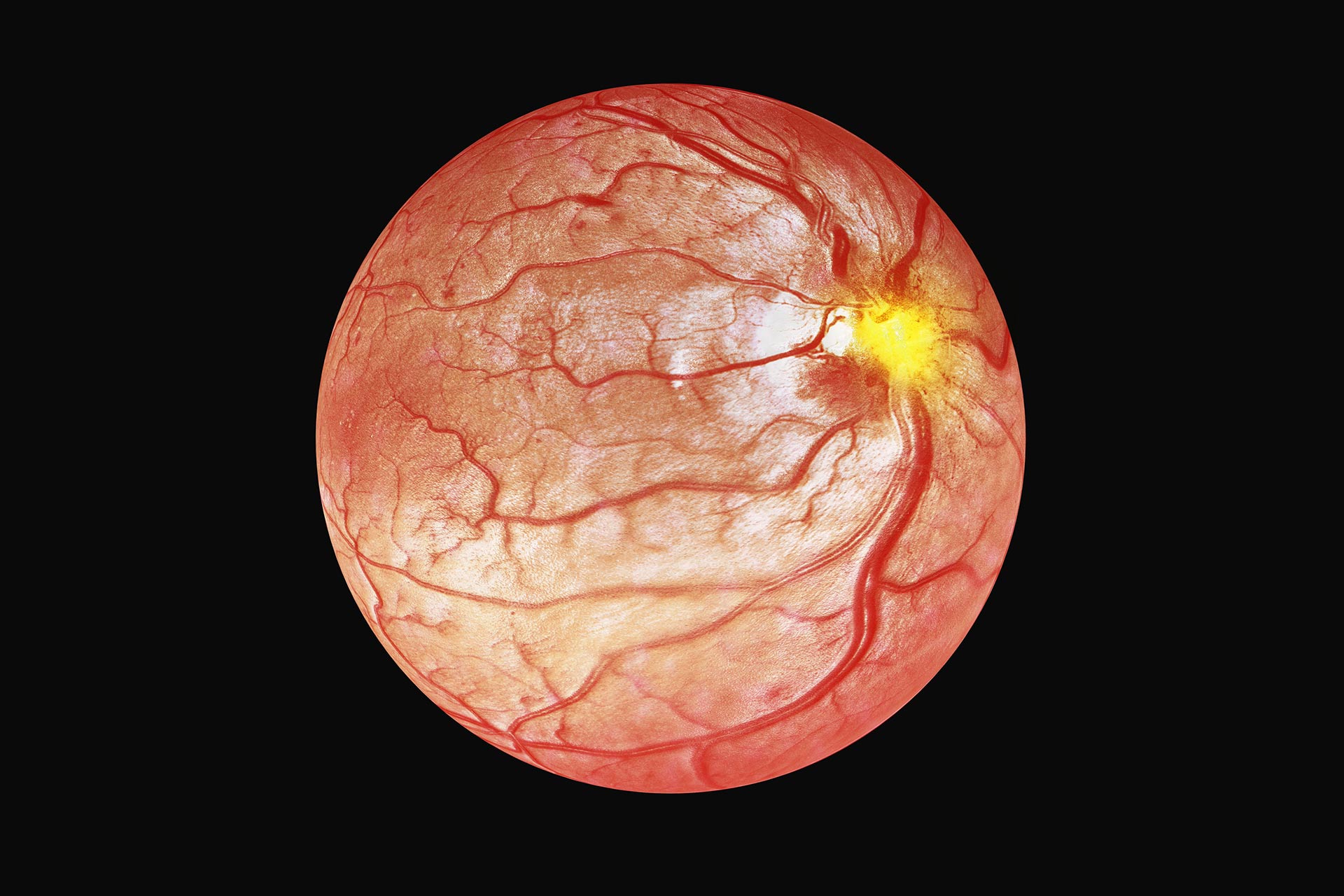
- Fundus Photography – we offer both traditional and wide-field fundus photography which is a non-invasive diagnostic procedure that provides photographs of the back of the eye to help determine the health of the optic nerve, vitreous, macula, retina and its blood vessels.
- Diagnostic Ocular Ultrasound - a technique in which a small ultrasound probe is used to make a virtual image of the eye with sound waves. Conditions such as a dense cataract or vitreous hemorrhage can prevent a direct view of the retina even with a dilated exam. When a view to the back of the eye is not possible, the ultrasound probe can produce an image of the entire eye. Ultrasound can also be used to diagnose and make measurements of a tumor within the eye.
- OCT Angiography (OCT-A) - a non-invasive technique that acquires wide field angiographic information without the use of dye unlike traditional fluorescein angiography (FA). Each three-dimensional (3D)scan set takes approximately six seconds to obtain. OCT angiography (OCT A) can image all layers of the retinal vasculature down to the capillary level unlike the traditional FA which provides a two-dimensional (2D) nature of the acquired images.
- Fluorescein Angiography - involves the injection of a small amount of vegetable-based dye through a patient's peripheral vein, usually the arm or hand. Shortly after, an ophthalmic photographer takes a series of time-dependent retinal photographs. The injected dye lights up the retina's intricate vascular network and helps pinpoint problem areas. In addition to the traditional Fluorescein Angiography, we offer state of the art, wide-field angiography which enables 80-90% improved visualization out in the far peripheral retina which is where a great deal of ocular pathology exists.
Treatment
If treatment of your eye problem is necessary, you and your physician will decide the most appropriate course of action. As a general rule, patients who receive treatment in a more timely fashion tend to fare better than those who delay evaluation by a retina specialist.
If it is discovered that you have wet or dry ARMD (aged-related macular degeneration), then you should monitor daily with the Amsler grid. We can supply this to you free of charge. Often, patients with subtle progression of macular disease will report new “waviness” or “missing areas” when monitoring their grid. One of our qualified or trained technicians, or nurse are always available to consult with you by phone.
Some types of treatment can be performed in our offices at the time of your visit while others are performed at the Baptist Eye Center on the surgery floor. Common procedures performed include:
- Intravitreal injections - "a procedure to place a medication directly into the space in the back of the eye called the vitreous cavity, which is filled with a gel-like fluid called the vitreous humor gel." The anti-VEGF drugs and steroids help to reduce fluid leakage associated with these disorders. Steroids, antibiotics, antiviral and antifungal medications can all be given via intravitreal injection. The procedure is usually performed by a trained retina specialist in the office setting. We currently offer all of the approved intravitreal drug therapies:
- Bevacizumab (Avastin)
- Ranibizumab (Lucentis)
- Aflibercept (Eylea)
- Dexamethasone (Ozurdex)
- Fluocinolone Acetonide (Iluvien)
- Fluocinolone acetonide (Yutiq)
- Vabysmo
The need for a repeat injection is determined during the clinical examination, often with the use of diagnostic testing such as optical coherence tomography (OCT)
and fluorescein angiography (FA).
Research is underway that will hopefully provide longer-acting treatments.
- Laser treatment - Laser therapy is used to treat a number of vitreoretinal conditions and can be done in our office as well as our satellite locations in many cases.
- Vitrectomy - A small cutting instrument is used to enter the eye through a 1 mm incision, and the vitreous gel is removed and replaced with a gas bubble. The advantage of this method is that all the debris, scar tissue and membranes on the retina can be removed, and the retina flattened at the time of surgery. A laser may be used to seal off the tears. This is not an office procedure and is scheduled in advance unless it is on an urgent basis.
Browse Our Website
Contact Information
Phone:
(501) 219-0900
Fax: (501) 312-4750
Baptist Eye Center
9800 Baptist Health Drive, Suite 200
Little Rock, AR 72205
Business Hours
- Mon - Fri
- -
- Sat - Sun
- Closed


Location
The Baptist Eye Center is located at EXIT 7 off I630, the 1st building on the right at the 3-Way stop sign